| Program C01-3 | Towards a new methodology of functional imaging for the central lymphatic system using an ultra-high-resolution SPECT |
|---|---|
| Principal Investigator | MIZUMA, Hiroshi (RIKEN) |
Immunity is the ability of the host to defend the body from external
invasions such as allergens and bacterial and viral infections. The
body's immune system responds to resist these invasions. Immune cells
play a key role in our body's protection. These cells mature in the bone
marrow and in the thymus, reaching the entire body via the blood and
lymphatic circulatory systems. The neuro-immune system is known to
have unique properties that are unlike the immune system in other
peripheral tissues. The neuro-immune system protects the brain by
restricting the passage of many molecules and cells between blood
vessels and brain parenchyma (blood-brain barrier). For centuries, it
was thought that the neuro-immune system lacked lymphatic vessels.
Using advanced fluorescent microscopic techniques, scientists
recently discovered lymphatic vessels in rodent and human
meninges. This discovery may open a new era, where additional
research will add to our understanding of the neuro-immune system. To
date, studies have revealed that the brain's lymphatic vessels play
important roles in both draining waste generated by neural activities
and interacting peripheral immune cells into brain
parenchyma. Changes in the brain's lymphatic system may be closely
related to aging and pathophysiological states, in particular,
Alzheimer's disease and multiple sclerosis.
The structural and functional properties of the brain's lymphatic
vessels have gradually been characterized. Yet, a way to
systematically visualize the central nervous system (CNS) lymphatic
vessels in live state has not been established. In this study, we
will develop a newer methodology for functional imaging of the CNS
lymphatic system using our in vivo imaging techniques on small
animals. To realize this, we will use single-photon emission computed
tomography (SPECT) with cadmium telluride (CdTe) detectors. This
technique had the potential to produce ultra-high spatial resolution
(≲100 μm) based on the cutting-edge technologies for observing
X- and gamma-rays in the universe. To visualize the CNS lymphatic
kinetics, we will test several SPECT tracers which are radiolabeled
to the macromolecules (e.g., ovalbumin) and T cells using Indium-111
(In-111), Gallium-67 (Ga-67), or Technetium-99m (Tc-99m).
By using our developed method, we will make a proposal for innovative
therapeutic strategies and/or diagnostic tools for neurologic
disorders and brain tumors associated with the dysfunction of the
neuro-immune system.
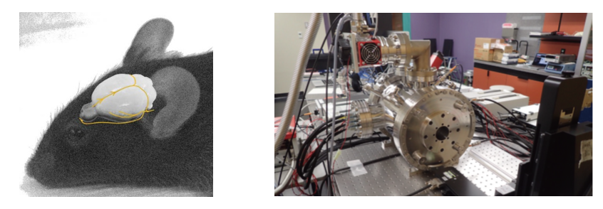
The schema of the CNS lymphatic tracts in a mouse (left). The
structural characterizations of CNS lymphatic vessels have been
revealed using advanced optical imaging techniques. To visualize
the brain’s lymphatic circulation, we use an ultra-high-resolution
SPECT scanner with radiolabeled macromolecules and immune cells
(right).
Members
- Principal Investigator
-
MIZUMA, Hiroshi

(RIKEN Center for Biosystems Dynamics Research (BDR))
- Research Collaborators
-
KANAYAMA, Yousuke (RIKEN BDR)
TAKEDA, Shin'ichiro (Kavli IPMU)
TAKAHASHI, Tadayuki (Kavli IPMU)
FUJII, Hirofumi (National Cancer Center)
Reference Materials
- H. Mizuma et al., “Establishment of in vivo brain imaging method in mice under conscious condition,” J. Nucl. Med. 51, 1068–1075 (2010).
- T. Kambe et al., “Differential regional distribution of phosphorylated tau and synapse loss in the nucleus accumbens in tauopathy model mice,” Neurobiol. Dis. 42, 404–414 (2011).
- Y. Hara et al., “Involvement of the septo-hippocampal cholinergic pathway in association with septal acetylcholinesterase upregulation in a mouse model of tauopathy,” Curr. Alzheimer Res. 14, 94–103 (2017).
- N. Nakai et al., “Serotonin rebalances cortical tuning and behavior linked to autism symptoms in 15q11-13 CNV mice,” Sci. Adv. 3, e1603001 (2017).