| Program C01-8 | Development of high resolution radionuclide imaging for peritoneal dissemination of gastric cancer |
|---|---|
| Principal Investigator | KUMAKURA, Yoshitaka (Saitama Medical University) |
Nuclear medicine is originally a non-invasive quantitative imaging
technique to visualize physiological and biochemical abnormalities of
human organs using small molecules such as glucose and amino
acids. This clinical technique uses radiolabeled pharmaceuticals
designed to accumulate in human organs of interest. Positron emission
tomography (PET) provides sectional images of radioactivity
distribution. Due to the recent improvement in spatial resolution,
preclinical PET with small animals has become a powerful tool for
development of candidate molecules for new drugs. These molecules
include tumor-specific monoclonal antibodies (mAbs). Currently, small
animal PET imaging is essential to evaluate mAb binding to grafted
tumors in immunocompromised nude mice. By incorporating therapeutic
radionuclides like α (or β) emitters with mAbs, highly
tumor-specific cell damages can be achieved. Here, PET imaging can
help optimize biodistribution of engineered mAb-based molecules. This
emerging therapeutic technique paired with molecular diagnostic
imaging (aka “theranostics”) is attracting a great deal of
attention to cure cancer patients. The bombardment of α particles
with high linear energy transfer (LET) can be delivered by mAbs to
surface antigens of tumor cells. This results in DNA double-strand
breaks, which ultimately induce apoptosis or necrosis of tumor
cells. We aim at development of novel theranostic strategies for
peritoneal dissemination of gastric cancer. As a therapeutic
α-emitter, Astatine-211 (half-life: 7.2 hours) is a promising
heavy halogen radionuclide, which can be produced by a
cyclotron. However, current imaging techniques for living small
animals are inadequate for visualization of 211At biodistribution, as
211At emits low energy X-rays (77–92 keV) and
scarce γ-rays with high energy (570 and 898 keV, <1%).
Thus, the high-resolution imaging counterpart
for 211At remains a challenge to establish a one-stop shop
approach for 211At theranostics. We expect that
novel 211At imaging techniques will be explored by multidisciplinary
collaborations, in comparison with the therapeutic outcomes after
targeted radionuclide therapy of 211At as well as the conventional
small animal PET images for disseminated peritoneal tumors of gastric cancer.
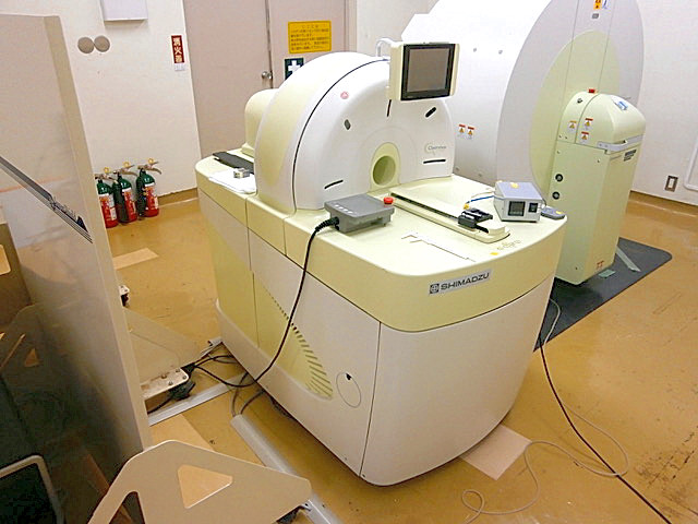 Fig 1. The preclinical PET scanner (Shimadzu corporation) installed at the Isotope Science Center, the University of Tokyo.
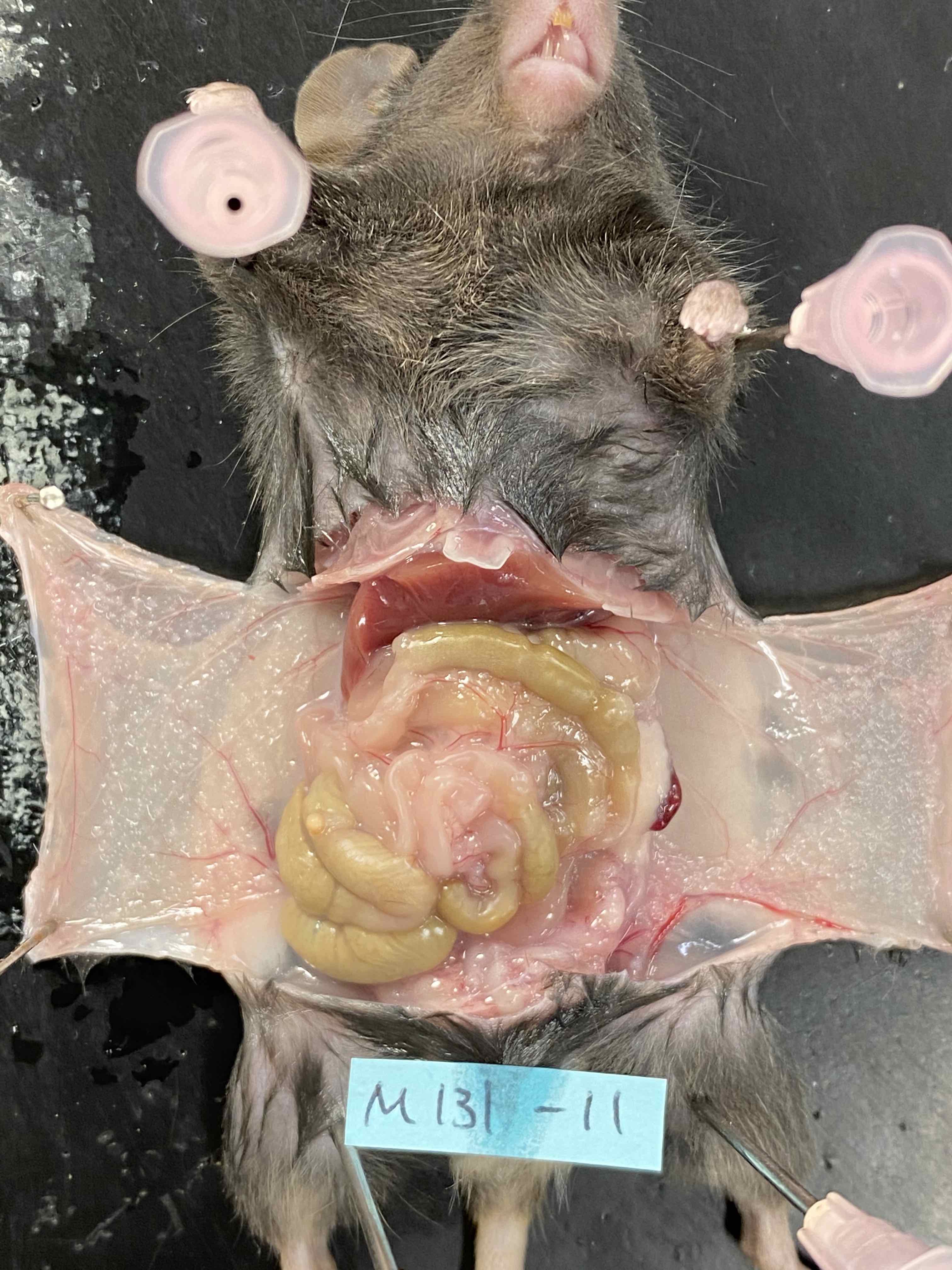 Fig 2. A representative image of an immunocompetent mouse with peritoneal dissemination of the gastric cancer cell line YTN16.
Members
- Principal Investigator
-
KUMAKURA, Yoshitaka
(Faculty of Medicine, Saitama Medical University)
- Research Collaborators
-
NOMURA, Sachiyo (The University of Tokyo)
WADA, Youichiro (The University of Tokyo)
AKIMITSU, Nobuyoshi (The University of Tokyo)
SUGIYAMA, Akira (The University of Tokyo)
HABA, Hiromitsu (RIKEN)
Reference Materials
-
M. Yamamoto ,S. Nomura ,A. Hosoi , et al., “Established gastric cancer cell lines transplantable into C57BL/6 mice show fibroblast growth factor receptor 4 promotion of tumor growth,” Cancer Sci. 109(5), 1480–1492 (2018). -
D. Fujimori ,J. Kinoshita ,T. Yamaguchi , et al., “Established fibrous peritoneal metastasis in an immunocompetent mouse model similar to clinical immune microenvironment of gastric cancer,” BMC Cancer 20(1), 1014 (2020). -
K. Nagaoka ,M. Shirai ,K. Taniguchi , et al., “Deep immunophenotyping at the single-cell level identifies a combination of anti-IL-17 and checkpoint blockade as an effective treatment in a preclinical model of data-guided personalized immunotherapy,” J. Immunother. Cancer 8(2), e001358 (2020). -
T. Kawato ,E. Mizohata ,Y. Shimizu , et al., “Structure-based design of a streptavidin mutant specific for an artificial biotin analogue,” J. Biochem. 157(6), 467–475 (2015). - 羽場宏光, 「理研における核医学治療用ラジオアイソトープの製造」, Drug Deliv. Syst. 35(2), 114–120 (2020). DOI: 10.2745/dds.35.114 .